Sonography
What is sonography in gastroenterology?
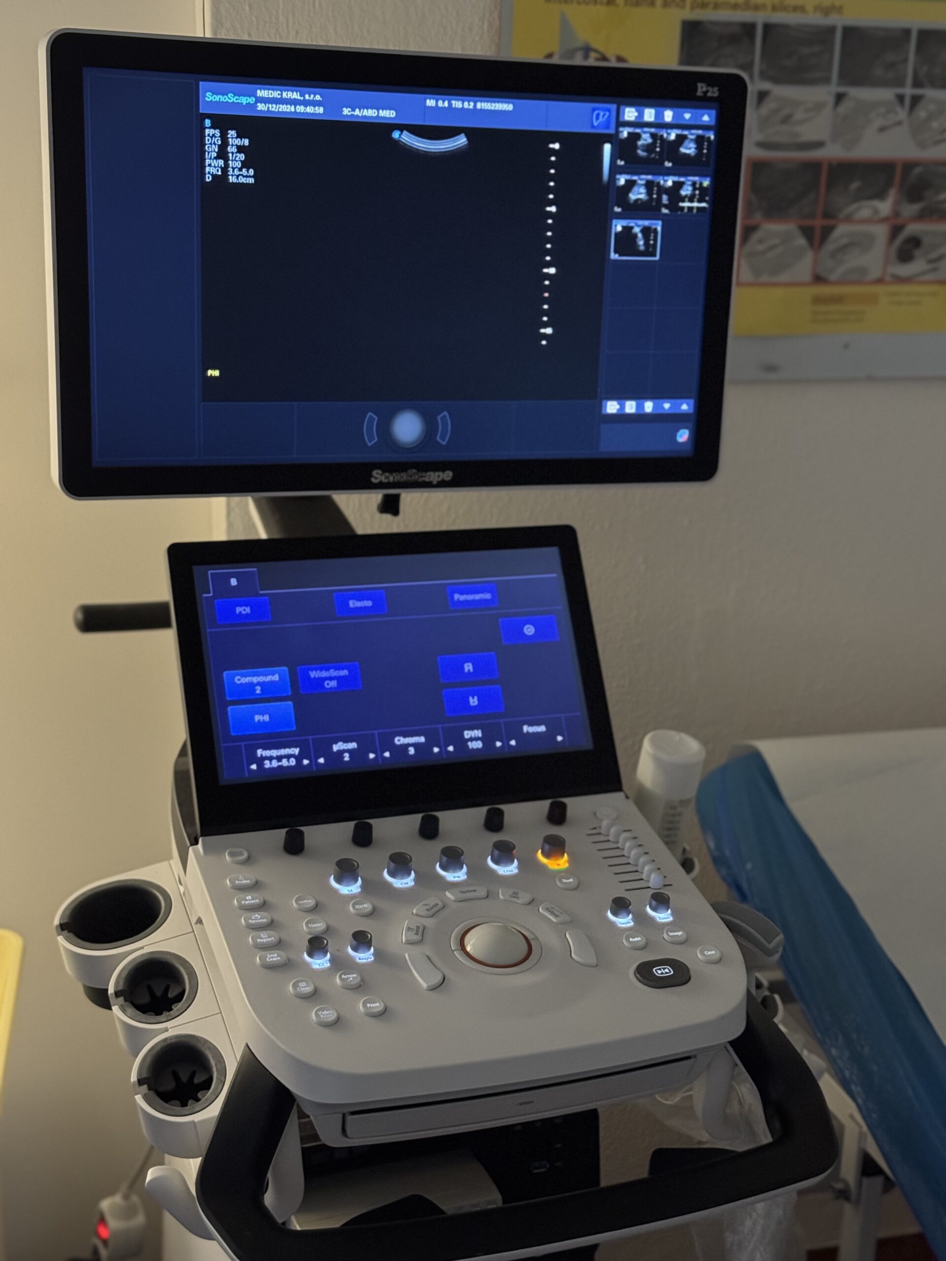
Sonography, also known as ultrasound examination, is a non-invasive diagnostic method that uses high-frequency sound waves to create images of internal organs and structures in the body. In gastroenterology, sonography is used to assess the condition of abdominal organs such as the liver, gallbladder, pancreas, spleen, stomach, intestines, and vascular structures in the area.
How does sonography work?
Ultrasound examination is completely painless and typically lasts 15–30 minutes. The patient lies on the examination table with the abdominal area exposed. The doctor applies a gel to the skin to allow smooth movement of the probe and improve the transmission of sound waves. The probe is the device that sends and receives sound waves, which bounce off internal organs and create images on a monitor.

Why is sonography performed?
Sonography in gastroenterology has wide diagnostic applications and is used to assess:
- Changes in size and structure of organs: such as liver enlargement (hepatomegaly) or spleen enlargement (splenomegaly).
- Presence of localized changes: like cysts, tumors, or abscesses.
- Condition of the gallbladder and bile ducts: detecting gallstones, cholecystitis (inflammation of the gallbladder), or bile duct obstruction.
- Condition of the pancreas: such as in cases of suspected acute or chronic pancreatitis or pancreatic tumors.
- Presence of fluid in the abdominal cavity: such as ascites (fluid accumulation).
- Blood circulation: using Doppler ultrasound to assess blood flow in blood vessels, for example, in the portal vein.
How to prepare for a sonography?
Preparation for abdominal sonography depends on the organs being examined. Generally, it is recommended to:
- Fast for at least 6 hours before the examination to reduce the amount of gas in the intestines and improve image quality.
- Avoid drinks that may cause gas formation, such as carbonated beverages.
- In some cases, the doctor may recommend medications to reduce bloating.
Is sonography painful?
Sonography is completely painless and very well tolerated by patients. Mild discomfort may occur when pressure is applied to the abdominal wall, especially in sensitive areas.
Significance and safety of sonography
Sonography is a safe procedure with no risk of exposure to ionizing radiation, which makes it suitable for repeated use without concerns about side effects. It is a key tool in gastroenterology for quick and accurate diagnosis of digestive system diseases. Due to its non-invasive nature, availability, and high diagnostic value, ultrasound plays an indispensable role in patient care.